Virtual Consultations

(831) 647-3900

Reconstructive Eyelid Surgery

What is Reconstructive Eyelid Surgery? Reconstructive eyelid surgery, also known as eyelid reconstruction, is a surgical procedure performed to excise and reconstruct the eyelid affected by cancerous growth. This procedure aims to remove the cancerous tissue and restore the functionality and aesthetic appearance of the eyelid. Eyelid cancer is a common occurrence, often associated with […]

Call Today For A Consultation!

(424) 354-2053

or schedule online by clicking here »

What is Reconstructive Eyelid Surgery?

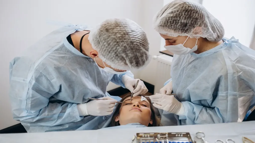
Reconstructive eyelid surgery, also known as eyelid reconstruction, is a surgical procedure performed to excise and reconstruct the eyelid affected by cancerous growth. This procedure aims to remove the cancerous tissue and restore the functionality and aesthetic appearance of the eyelid.

Eyelid cancer is a common occurrence, often associated with prolonged sun exposure. In fact, it is estimated that about 90% of eyelid cancers are caused by excessive exposure to the sun’s harmful ultraviolet (UV) rays. This makes the eyelids particularly vulnerable to developing cancerous lesions.

Reconstructive Eyelid Surgery

There are four principal factors related to reconstructive eyelid surgery. First, the excision of the cancerous tissue involves careful removal of the affected eyelid area to ensure complete eradication of the cancer. Second, reconstruction techniques are employed to restore the eyelid’s form and function. The goal is to reconstruct the eyelid in a way that preserves its ability to protect the eye and maintain adequate tear production. Third, special attention is given to the aesthetic outcome, aiming to achieve a natural appearance that blends harmoniously with the surrounding facial features. Finally, sun protection measures and education on sun safety are vital to preventing the recurrence of eyelid cancer and ensuring the long-term success of the reconstruction.

Upper Eyelid Reconstructive Surgery

Upper Eyelid Reconstructive Surgery

Upper eyelid reconstructive surgery is a specialized procedure aimed at correcting various issues and restoring the natural appearance and functionality of the upper eyelid.Whether due to trauma, congenital abnormalities, or age-related changes, an upper eyelid that is droopy, disfigured, or impaired can negatively impact both physical appearance and visual acuity. This surgical intervention involves meticulously reshaping, restoring, or repairing the upper eyelid, ensuring optimal aesthetics and, in many cases, improved vision.

By addressing and resolving any functional or cosmetic concerns, upper eyelid reconstructive surgery can significantly enhance a patient’s quality of life, boosting self-confidence and overall well-being. By exploring the following headings, we will delve into the different aspects of this procedure, including candidacy, techniques, recovery, and potential risks and complications, providing a comprehensive understanding of upper eyelid reconstructive surgery.

Pretarsal Defects of the Upper Eyelid

Pretarsal defects of the upper eyelid can result from various causes, such as trauma, tumors, or previous surgical procedures. Several approaches can be utilized to address these defects, depending on their size and location. This article will discuss the different methods commonly used, including full-thickness skin grafting, wedging and primary closure, Tenzel semicircular flap with lateral canthal tendon division, crisscross canthoplasty for defects involving both eyelids, and the use of a lid switch flap for larger defects.

Full-thickness skin grafting is an effective approach for larger pretarsal defects. It involves removing a full-thickness piece of skin from a donor site, typically the upper eyelid or postauricular area, and transplanting it onto the defect. This technique provides good function and cosmesis but does require an additional surgical site.

For smaller defects, wedging and primary closure can be utilized. This technique involves removing a triangular-shaped wedge of tissue from the defect and then bringing the remaining edges together for closure. This approach often provides excellent aesthetic results and preserves eyelid integrity.

The Tenzel semicircular flap with lateral canthal tendon division is used for moderate-sized defects. In this technique, a semicircular flap is created adjacent to the defect, and the lateral canthal tendon is divided to allow for flap advancement. This approach helps maintain eyelid tension and is particularly useful for situations where primary closure is not feasible.

In cases where both eyelids are affected, a crisscross canthoplasty approach can be used. This technique involves creating a triangular flap on each eyelid and interdigitating their tips to achieve closure. It is particularly useful for larger, more complex defects and allows for improved function and symmetry.

Lastly, for larger defects, a lid switch flap can be employed. This technique entails transposing a healthy portion of the upper eyelid to cover the defect. It preserves eyelid function and contour but may require additional reconstructive procedures.

Preseptal Defects of the Upper Eyelid

Preseptal defects refer to any abnormalities or damage to the tissues surrounding the eyeball but not involving the deeper structures like the orbit or eyelid muscle. When repairing preseptal defects, it is important to consider upper lid fold defects and the role of location and dermatochalasis.

Upper lid fold defects occur when the natural contour of the upper eyelid fold is disrupted due to trauma, surgery, or congenital issues. Repairing upper lid fold defects requires restoring the natural contour and ensuring proper eyelid function. Techniques used for repair include skin grafts or local flaps, depending on the extent and location of the defect. Skin grafts involve taking healthy skin from another area of the body and transplanting it to the affected area. Local flaps involve transferring neighboring tissue to reconstruct the defect. The choice between these techniques depends on the surgeon’s evaluation of the defect and the patient’s individual circumstances.

Location of the defect is crucial as it determines the approach for repair. Medial defects, which occur towards the inner corner of the eye, are best repaired using local flaps or skin grafts rather than primary closure. This is because the surrounding tissue in this area is limited, and using local flaps or grafts provides better aesthetic and functional outcomes. On the other hand, lateral defects, closer to the outer corner of the eye, can be more forgiving as there is often excess skin in this area. This allows for primary closure without significant loss of function or aesthetic compromise.

When performing upper blepharoplasty, proper incision design and execution are crucial for successful outcomes. The surgeon must carefully mark the crease, which is the natural fold of the upper eyelid, to ensure symmetry and proper contouring. Maintaining the three arcs of the upper eyelid-brow subunit, which include the eyebrow arch, supraorbital arc, and upper lid fold, is necessary to create a natural and aesthetically pleasing appearance.

Medial Canthal Defects

Medial canthal defects refer to damage or deformities in the area near the inner corner of the eye. These defects can occur due to trauma, surgery, or congenital abnormalities. Fortunately, there are several techniques available to address medial canthal defects and restore the normal appearance and function of the eye.

One technique commonly used is the full-thickness skin graft. This involves harvesting a piece of skin from a donor site, such as the upper eyelid or the posterior auricular region, and grafting it onto the defect. The graft is secured in place with sutures, and over time, it integrates with the surrounding tissues, providing a stable and durable repair.

Another technique is the use of glabellar flaps. These flaps are created by mobilizing skin and underlying tissue from the forehead region and rotating it to cover the defect. Glabellar flaps are particularly useful for larger defects or when there is limited local tissue available for grafting.

Forehead flaps are another option for addressing medial canthal defects. Similar to glabellar flaps, forehead flaps involve using adjacent tissue from the forehead to reconstruct the area around the eye. These flaps offer good vascularity and can be an effective solution for complex defects.

In some cases, secondary intention healing may be chosen instead of surgical reconstruction. This involves allowing the defect to heal on its own without intervention. While this technique may not provide the same cosmetic outcomes as grafts or flaps, it can be a suitable option for smaller defects or in instances where surgery may not be appropriate.

When addressing medial canthal defects, it is crucial to fix the medial canthal attachments to the periosteum or bone using heavy permanent sutures. These sutures help stabilize the surrounding tissues and prevent complications such as ectropion (outward turning of the eyelid) or entropion (inward turning of the eyelid). Additionally, other methods like wiring or miniplates can be used to secure the attachment.

One important consideration in medial canthal defect reconstruction is the potential for lacrimal injury. The lacrimal system, responsible for tear production and drainage, runs through the medial canthal area. In cases where there is significant injury to the lacrimal system, additional reconstructive procedures may be required. This can involve canaliculostomy, which creates a new connection between the eye and nasal cavity, and medial canthoplasty, which helps restore the integrity of the canthal attachments and maintain proper tear drainage.

Medial Canthal Defects

Lower Eyelid Reconstructive Surgery

Lower Eyelid Reconstructive Surgery is a surgical procedure aimed at correcting various issues affecting the lower eyelid. From congenital defects to trauma-related injuries and the natural aging process, this surgery can address a range of concerns. The procedure involves the careful removal or repositioning of excess skin, fat, and muscle, in order to restore a more youthful and balanced appearance to the lower eyelid. Additionally, it can correct sagging, drooping, or malpositioned lower eyelids that may be hindering proper tear drainage. Whether performed for functional or cosmetic reasons, lower eyelid reconstructive surgery can offer significant improvements in both appearance and eye health.

Ectropion

Lower Eyelid Reconstructive Surgery

Ectropion is a condition characterized by the abnormal outward turning and drooping of the lower eyelid. This can occur due to various causes, such as age-related changes in the cartilage of the lower eyelid or as a result of facial nerve weakness or scarring.

Symptoms of ectropion may include excessive tearing, dryness and irritation of the eye, sensitivity to light, redness, and a gritty or sandy feeling in the eye. The outward turning and sagging of the lower eyelid can cause the inner surface of the eyelid to become exposed, leading to dryness and inflammation of the conjunctiva.

Surgical repair options are available to correct ectropion and restore proper function and appearance to the eyelid. The specific procedure used will depend on the underlying cause and severity of the condition.

One common surgical approach involves tightening the lower eyelid and restoring its normal position by removing a small section of skin and tightening the underlying tissues. In cases where facial nerve weakness is the underlying cause, additional procedures may be necessary to address the underlying nerve dysfunction.

Aging can have significant effects on the cartilage of the lower eyelid, leading to its drooping and outward turning. Over time, the cartilage can weaken and lose its elasticity, causing the lower eyelid to lose its ability to maintain proper alignment. This can result in the lid sagging downward and turning outward, a condition known as involutional ectropion.

Pretarsal Defects of the Lower Eyelid

Pretarsal defects of the lower eyelid are complex anatomical challenges often arising from trauma, surgical excision of malignancies, or congenital anomalies. These defects involve the pretarsal region, which is the part of the lower eyelid immediately adjacent to the eyelashes. Given the intricate structure of the lower eyelid, which includes a delicate balance of skin, muscle, and connective tissue, the repair of pretarsal defects requires meticulous planning and precise surgical techniques to restore both function and aesthetics.

The primary goals in managing pretarsal defects include preserving eyelid mobility, ensuring adequate coverage of the eyeball, and achieving an aesthetically pleasing result. Several reconstructive techniques can be employed, ranging from local flaps and grafts to more complex procedures such as tarsoconjunctival flaps or full-thickness grafts from the upper eyelid. The choice of technique depends on the size and location of the defect, as well as the overall health and needs of the patient.

Successful reconstruction not only restores the protective function of the eyelid but also addresses the aesthetic concerns, which are critical given the eyelid’s prominent role in facial expression. Complications such as ectropion, entropion, or improper eyelid closure can significantly impact the patient’s quality of life, making the expertise and experience of the surgeon paramount in achieving optimal outcomes. Advances in microsurgical techniques and a deeper understanding of eyelid anatomy continue to enhance the effectiveness of treatments for pretarsal defects, offering patients improved functional and cosmetic results.

Preseptal Defects of the Lower Eyelid

Preseptal defects of the lower eyelid are typically less complex than pretarsal defects but still present significant challenges in terms of both function and aesthetics. The preseptal region, located anterior to the orbital septum, consists primarily of skin and subcutaneous tissue, playing a crucial role in the overall contour and integrity of the lower eyelid. Defects in this area can result from trauma, surgical excision of skin cancers, infections, or congenital conditions.

The primary objective in managing preseptal defects is to restore the natural appearance and function of the eyelid while preventing complications such as eyelid retraction or asymmetry. Treatment options vary based on the size and depth of the defect, ranging from primary closure for small defects to local flaps, such as V-Y advancement flaps or rotation flaps, for more extensive tissue loss. Skin grafts, either full-thickness or split-thickness, may be necessary when local tissue is insufficient to cover the defect adequately.

In addition to ensuring a cosmetically pleasing result, the reconstruction must preserve the protective function of the eyelid, which includes maintaining proper lubrication and protection of the ocular surface. Careful consideration of the vascular supply, tissue characteristics, and potential for scarring is essential in planning the reconstruction to achieve optimal outcomes. Advances in reconstructive techniques and an in-depth understanding of eyelid anatomy have significantly improved the prognosis for patients with preseptal defects, enabling the restoration of both form and function with minimal morbidity.

Eyelid–cheek Junction

The eyelid-cheek junction is a critical area of the face that can be prone to various defects. These defects can result from aging, trauma, or surgical procedures. When defects occur at the eyelid-cheek junction, it can lead to a loss of volume in this area, resulting in a sunken appearance. In such cases, volume restoration becomes necessary to achieve a natural and youthful look.

There are several options for reconstructing defects at the eyelid-cheek junction. One option is the use of injectable fillers such as hyaluronic acid. These fillers can be injected into the area to restore volume and smooth out any depressions. Another option is fat grafting, where fat is harvested from one part of the body and injected into the eyelid-cheek junction.

For larger defects, raising the cervicofacial flap in the deep-plane can be beneficial. This technique involves elevating the skin and underlying fat and muscle in the deep-plane, which provides better blood supply and reduces the risk of complications. This approach allows for larger defects to be reconstructed effectively.

Considerations for bony support at the eyelid-cheek junction are important as well. The orbital rim provides support for the lower eyelid, and any defects in this area can lead to functional and aesthetic issues. Reconstructing the orbital rim can be done using various methods, including the use of implants, autologous grafts, or allografts.

Functional Eyelid Reconstructive Surgery

Functional eyelid reconstructive surgery is a specialized procedure designed to improve the functioning and appearance of the eyelids. Whether due to injury, disease, or congenital defects, issues with the eyelids can cause significant discomfort and impact a person’s quality of life. However, thanks to advancements in medical technology and surgical techniques, patients can now undergo reconstructive surgery to restore normal eyelid function and regain their confidence.

Eyelids play a crucial role in protecting the eyes, spreading tears evenly across the surface, and controlling the amount of light that enters the eye. When these functions are compromised, individuals may experience a range of problems such as dry eyes, excessive tearing, difficulty closing or opening the eyes, and even vision impairment.

Functional eyelid reconstructive surgery addresses these issues by correcting abnormalities or damage to the eyelids. The procedure involves meticulous surgical techniques that aim to preserve the natural form and function of the eyelids while achieving an aesthetically pleasing outcome. Surgeons who specialize in this field have extensive knowledge of the complex anatomy of the eyelids and employ innovative approaches to meet the unique needs of each patient.

There are various reasons why a person may require functional eyelid reconstructive surgery. Traumatic injuries, such as those caused by an accident or a burn, can result in eyelid deformities and restrict normal eyelid movement. Additionally, certain medical conditions like Bell’s palsy, cancer, or facial nerve paralysis can lead to eyelid dysfunction. Congenital defects, such as ptosis (droopy eyelids) or ectropion (outwardly turned eyelids), may also necessitate surgical intervention.

During the consultation phase, the surgeon will evaluate the patient’s condition, medical history, and desired outcomes. In many cases, the surgeon will work closely with other specialists, to develop a comprehensive treatment plan. They will take into consideration factors such as the severity of the condition, the patient’s overall health, and any previous surgeries.

The actual surgery can vary depending on the specific needs of the patient. It may involve procedures such as eyelid reconstruction, eyelid tightening, eyelid lift, or eyelid repositioning. The surgeon will utilize various techniques, such as skin grafts, tissue rearrangement, or the use of synthetic materials, to restore the functionality and appearance of the eyelids. The goal is to ensure that the eyelids can move freely, protect the eye adequately, and achieve symmetry with the other eyelid.

Recovery time will depend on the complexity of the surgery, but patients can generally expect some swelling, bruising, and discomfort in the immediate post-operative period. The surgeon will provide detailed aftercare instructions, which may include the use of eye drops, cold compresses, and regular follow-up appointments. Over time, as the swelling subsides and the tissues heal, patients will begin to notice a significant improvement in eyelid function and overall appearance.

Functional eyelid reconstructive surgery not only restores normal eyelid function but also has a significant impact on a person’s emotional well-being. Patients who have undergone this procedure often report enhanced self-confidence, improved vision, and an overall improvement in their quality of life.

Functional Eyelid Reconstructive Surgery

Corneal Abrasion

Corneal abrasion is a common eye injury that can cause discomfort and affect vision. It occurs when the clear, protective outer layer of the eye, known as the cornea, is scratched or scraped. While corneal abrasions can occur in various ways, such as from foreign objects, contact lenses, or even harsh chemicals, the symptoms and treatment remain consistent.

The symptoms of a corneal abrasion can vary depending on the severity of the injury. Common signs include eye pain, watering, redness, a sensation of something in the eye, sensitivity to light, and blurred vision. It’s essential to seek medical attention if you suspect you have a corneal abrasion, as improper treatment or neglecting the injury can lead to complications.

When a corneal abrasion is confirmed, the initial step in treatment is pain management. Eye drops or ointments may be prescribed to alleviate discomfort and reduce the risk of infection. It’s crucial to rest the affected eye, avoiding activities that may worsen the injury, such as rubbing or scratching. In some cases, an eye patch or soft contact lens may be recommended to protect the cornea and promote healing.

It’s important to note that the cornea has fantastic self-healing abilities. Most corneal abrasions heal within a few days to a week, depending on the severity. During this time, it’s essential to follow the doctor’s instructions carefully and refrain from wearing contact lenses or applying eye makeup. Any activity that may increase eye strain or introduce foreign substances should be avoided to prevent further complications.

Complications arising from corneal abrasions are quite rare but can occur if the injury is not appropriately managed.

Corneal Abrasion

These complications include corneal infections, corneal ulcers, and scarring. To minimize these risks, it’s crucial to attend follow-up appointments as scheduled and report any worsening symptoms or concerns to the healthcare provider promptly.

Prevention is always better than cure when it comes to corneal abrasions. Some simple measures can greatly reduce the risk of this injury. Wearing protective eyewear, such as safety goggles, when engaging in activities like woodworking, sports, or other environments where foreign objects may pose a risk. Properly caring for and cleaning contact lenses is also crucial, as improper hygiene can lead to corneal abrasions and other eye infections.

Dry Eye and Chemosis

Dry eye and chemosis are two conditions that can affect the eyes, causing discomfort and affecting vision. While both conditions are separate, they often occur together and share similar causes and treatments. Understanding these conditions is crucial to receive proper treatment and alleviate symptoms.

Dry eye syndrome, also known as keratoconjunctivitis sicca, occurs when the eyes do not produce enough tears or tears evaporate too quickly. Dry eye can result from various factors, such as age, hormonal changes, certain medications, environmental conditions, or underlying health conditions like diabetes or autoimmune disorders. Common symptoms of dry eye include redness, itching, a gritty sensation, blurred vision, and increased sensitivity to light.

Chemosis, on the other hand, refers to the swelling of the conjunctiva, the clear tissue that covers the white part of the eye. It is typically caused by the inflammation of blood vessels in the conjunctiva, resulting from allergies, infections, or irritants like dust, smoke, or chemicals. Symptoms of chemosis include swelling, redness, excessive tearing, and a feeling of irritation or foreign body sensation in the eye.

The link between dry eye and chemosis lies in inflammation. Dry eye can lead to inflammation of the conjunctiva, which can trigger chemosis. Additionally, chemosis itself can contribute to dry eye symptoms by disrupting the tear film, causing further discomfort and dryness.

Treatment for dry eye and chemosis often involves managing inflammation and optimizing tear production. Artificial tears, which mimic the composition of natural tears, can provide lubrication and relief for dry eye symptoms. Additionally, doctors may prescribe anti-inflammatory eye drops or ointments to reduce inflammation and promote healing.

For more severe cases, punctal plugs may be recommended. These tiny plugs are inserted into the tear ducts to block drainage, helping tears to stay on the eye’s surface for a longer time. In some cases, oral medication or steroid eye drops may be prescribed to address underlying conditions or manage severe inflammation.

Self-care measures can also aid in managing dry eye and chemosis symptoms. Avoiding prolonged screen time, taking regular breaks, using a humidifier, and protecting the eyes from dry or windy environments can provide relief. Wearing sunglasses outdoors and avoiding known allergens or irritants can also help reduce chemosis symptoms.

Lagophthalmos

Lagophthalmos is a medical condition characterized by the inability to fully close one’s eyelids. The term originates from the Greek word “lagos,” which means hare, and “ophthalmos,” meaning eye. This analogy is used because rabbits and some other animals can have a visible portion of their eyes when their eyelids are partially closed, similar to individuals with lagophthalmos. This condition can be quite distressing for those who experience it, as it can cause various eye problems and affect overall visual health.

The causes of lagophthalmos can vary, with several underlying factors contributing to the condition. One common cause is facial nerve paralysis, often resulting from conditions such as Bell’s palsy or trauma, affecting the nerve responsible for controlling eyelid movement. Other causes include congenital malformations, eyelid surgery complications, and certain medical conditions, such as thyroid eye disease and facial nerve tumors. Regardless of the cause, the primary concern with lagophthalmos is the exposure of the cornea, which can lead to dryness, irritation, and potentially serious complications if left untreated.

One of the key problems associated with lagophthalmos is the development of dry eye syndrome. Since the eyelids are unable to fully close, tears evaporate more quickly, leading to insufficient lubrication of the eye’s surface. This can cause discomfort, foreign body sensation, redness, and even visual disturbances. Moreover, a compromised cornea is at a higher risk of developing corneal ulcers and infections, which, if not adequately managed, can lead to vision loss.

Fortunately, various management techniques exist to alleviate the symptoms and address the underlying causes of lagophthalmos. The primary goal is to ensure adequate lubrication and protection of the eye to prevent dryness and potential corneal damage. This can be achieved through a range of strategies, including the use of artificial tears, gels, or ointments to provide extra moisture. In severe cases, the application of eye patches or moisture chamber glasses can be helpful, as they help create a protective barrier against environmental factors and help retain moisture.

Surgical interventions may also be considered in certain instances, particularly for cases involving facial nerve paralysis or structural abnormalities. Procedures such as tarsorrhaphy, a surgical technique that partially sews the eyelids together, can be performed to reduce exposure and promote proper lubrication. Additionally, eyelid weights or gold implants may be used to aid in eyelid closure.

It is crucial for individuals with lagophthalmos to seek timely medical attention and follow a comprehensive treatment plan to prevent significant eye-related complications. Regular consultations with oculofacial plastic surgeon and eye care professionals can ensure proper monitoring and adjustment of treatment strategies as necessary.

Eyelid Reconstructive Surgery Summary

Eyelid reconstructive surgery is a medical procedure that aims to correct abnormalities or damage to the eyelids. This type of surgery is often used to address conditions such as droopy eyelids, ptosis, ectropion, and entropion.

Droopy eyelids, also known as ptosis, can occur due to age, genetics, or muscle weakness. This condition can not only affect a person’s appearance but also interfere with their vision. Eyelid reconstructive surgery can correct ptosis by repositioning the eyelid muscles and removing excess skin or fat.

Ectropion and entropion are conditions in which the eyelids turn outward or inward, respectively. These conditions can cause irritation, redness, tearing, and discomfort. Eyelid reconstructive surgery can correct these issues by adjusting the position and tension of the eyelid muscles, restoring normal eyelid functionality.

The procedure itself involves making small incisions along the natural creases of the eyelids. The surgeon will then carefully tighten or reposition the muscles, remove excess tissue, and close the incisions with sutures. The surgery is typically performed under local anesthesia, and patients can expect a relatively short recovery period.

Eyelid reconstructive surgery not only improves the function of the eyelids but also enhances the overall aesthetic appearance of the eyes. Many people opt for this surgery to achieve a more youthful and rejuvenated look. The procedure can also address asymmetry or restore symmetry to the eyelids, creating a more balanced and harmonious appearance.

Before undergoing eyelid reconstructive surgery, a consultation with a board-certified oculoplasty surgeon like Dr. Bidar is essential. The surgeon will evaluate the individual’s specific concerns, medical history, and overall health to determine if they are a suitable candidate for the procedure. They will also discuss the potential risks, benefits, and expected outcomes to ensure realistic expectations.

CONTACT US