Virtual Consultations

(831) 647-3900

Orbital Reconstruction

What is Orbital Reconstruction? Orbital reconstruction refers to a surgical procedure aimed at restoring the external and internal orbital anatomy to its original or premorbid form. The goal of this procedure is to correct deformities or abnormalities in the orbital region caused by trauma, congenital anomalies, or previous surgeries. During orbital reconstruction, the surgeon works […]

Call Today For A Consultation!

(424) 354-2053

or schedule online by clicking here »

What is Orbital Reconstruction?

Orbital reconstruction refers to a surgical procedure aimed at restoring the external and internal orbital anatomy to its original or premorbid form. The goal of this procedure is to correct deformities or abnormalities in the orbital region caused by trauma, congenital anomalies, or previous surgeries.

During orbital reconstruction, the surgeon works to repair and reposition injured soft tissues within the orbit. This may involve the use of bone grafts, implants, or other materials to reconstruct the orbital walls and restore the integrity of the orbit. The ultimate objective is to optimize aesthetics, preserve eye function, and improve overall quality of life for the patient.

What is Orbital Reconstruction

Computer-assisted planning and intraoperative navigation/imaging play a crucial role in enhancing postoperative results. By using advanced imaging techniques and computer software, surgeons can accurately assess the extent of the orbital defect and plan the surgical intervention accordingly. This technology allows for precise preoperative measurements and simulations, ensuring optimal outcomes and minimizing the risk of complications. During the surgery, real-time intraoperative navigation helps guide the surgeon’s movements and ensures accurate placement of implants or grafts.

Secondary orbital reconstructions are commonly indicated in cases of globe malposition, binocular diplopia (double vision), and impaired eye motility. These complications may arise due to inadequate initial reconstruction or postoperative changes over time. In such cases, a secondary reconstruction is necessary to address the functional and aesthetic issues faced by the patient.

Overall, orbital reconstruction is a complex surgical procedure that aims to restore the normal structure and function of the orbit. The utilization of computer-assisted planning and intraoperative navigation greatly assists in achieving favorable outcomes, while secondary reconstructions are often required to address specific complications that arise postoperatively.

Types of Orbital Fractures

Types of Orbital Fractures

Orbital fractures refer to the breakage or damage to the bones located around the eye socket. These fractures can occur due to a variety of reasons, such as trauma from accidents, sports injuries, or even surgical interventions. Understanding the different types of orbital fractures is crucial for accurate diagnosis and appropriate treatment. This article aims to provide an overview of the various classifications of orbital fractures, including blowout fractures, tripod fractures, zygomaticomaxillary complex fractures, and orbital rim fractures.

By familiarizing ourselves with these different types, we can better comprehend the nature and extent of the injury, facilitating the intervention required for proper healing and restoration of function.

Orbital Roof Fracture

An orbital roof fracture is a relatively rare type of craniofacial fracture that involves the breakage of the thin, delicate bony structure that forms the roof of the eye socket or orbit. It is estimated that orbital roof fractures account for less than 2% of all facial fractures. The rarity of this fracture can be attributed to the protective anatomical position of the orbit in the skull.

The most common causes of orbital roof fractures are adjacent trauma to the skull. This can occur due to incidents such as motor vehicle accidents, falls, sports injuries, or direct blows to the face. The orbital roof can also be damaged by hydraulic forces transmitted from other facial fractures, particularly those involving the frontal bone or the medial and lateral walls of the orbit.

In addition to the fracture itself, orbital roof fractures can be associated with various intracranial injuries. These can include intracerebral hemorrhage, epidural hematoma, subdural hematoma, and traumatic brain injury. The severity of these intracranial injuries, as well as the presence of other associated facial fractures, are important factors in determining the need for surgical intervention.

Clinical indications for surgical intervention in orbital roof fractures include significant displacement of the fractured fragments, significant intracranial injuries, worsening or persisting neurologic deficit, severe pain, visual disturbances, double vision, or orbital entrapment. Prompt surgical intervention is crucial to prevent further complications and to restore the normal anatomy and function of the eye socket.

Zygomaticomaxillary Orbital Rim Fractures

Zygomaticomaxillary orbital rim fractures, also known as tripod fractures, occur due to high-impact trauma to the zygomaticomaxillary complex. This complex consists of the zygomatic bone, which forms the cheekbone, and the maxilla, which forms the upper jaw. These fractures are characterized by fractures involving three important structures: the zygomatic arch, the infraorbital rim, and the lateral orbital wall.

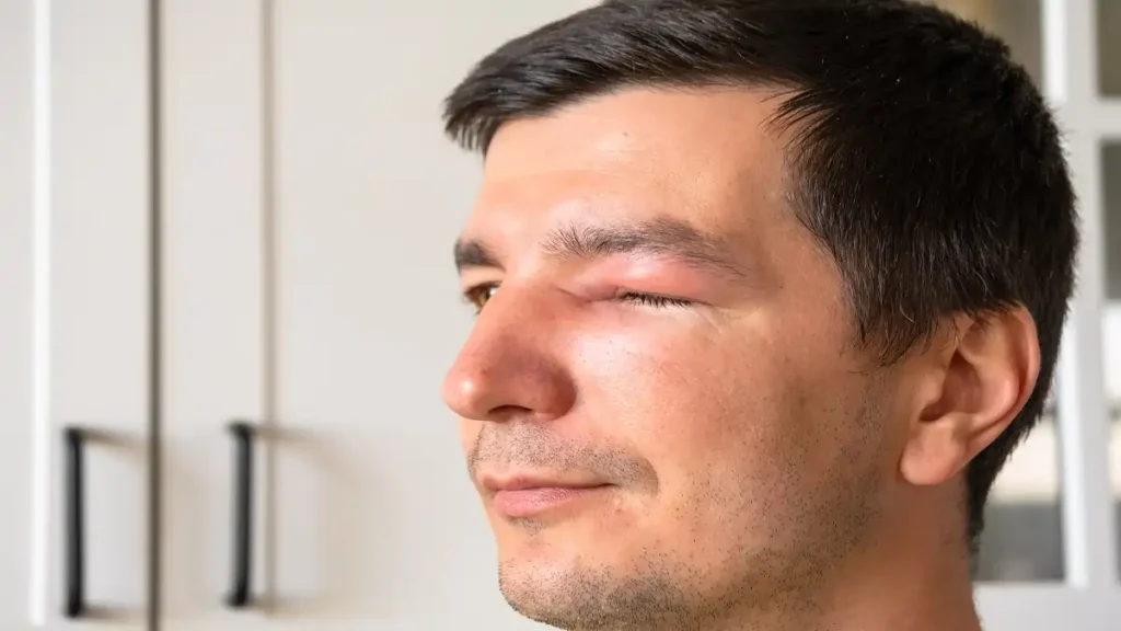
The causes of zygomaticomaxillary orbital rim fractures are usually blunt force trauma, such as from motor vehicle accidents, falls, or physical assaults. These fractures often result in visible facial deformity, periorbital ecchymosis (bruising around the eye), diplopia (double vision), infraorbital nerve anesthesia (numbness of the cheek), and restricted eye movement.

Repairing zygomaticomaxillary orbital rim fractures typically involves surgical interventions. The approaches used for repair vary depending on the complexity and location of the fracture. Often, a combination of intraoral and extraoral incisions are made to access the fracture site. Miniplates and screws are commonly used for stable fixation. These devices are typically made of titanium and provide rigid fixation to allow for proper bone healing.

The miniplates are placed along the zygomaticomaxillary complex and secured with screws. This technique ensures anatomical reduction and stable fixation, which leads to improved healing and restoration of facial symmetry and function.

Orbital Floor Fractures

Orbital floor fractures are a common type of facial fracture that involve the bones surrounding the eye socket. There are two main types of orbital floor fractures: blow out fractures and trap door fractures.

Blow out fractures occur when a blunt force trauma, such as a punch or a ball hitting the eye, causes the floor of the orbit to fracture. These fractures typically result in the herniation of the orbital contents into the maxillary sinus. The trapped air or fluid in the sinus can cause pressure and lead to complications such as diplopia (double vision), enophthalmos (sunken eye), or hypoesthesia (numbness) in the cheek and upper lip.

On the other hand, trap door fractures are a more specific subtype of blow out fractures. In trap door fractures, a segment of the orbital floor fractures and hangs like a trap door, blocking the normal movement of the orbital contents. This can lead to more severe complications such as inferior rectus muscle entrapment, which causes restricted eye movements and diplopia. Prompt surgical intervention is usually required with these fractures to release the entrapped muscle and restore normal eye function.

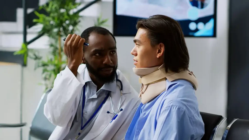
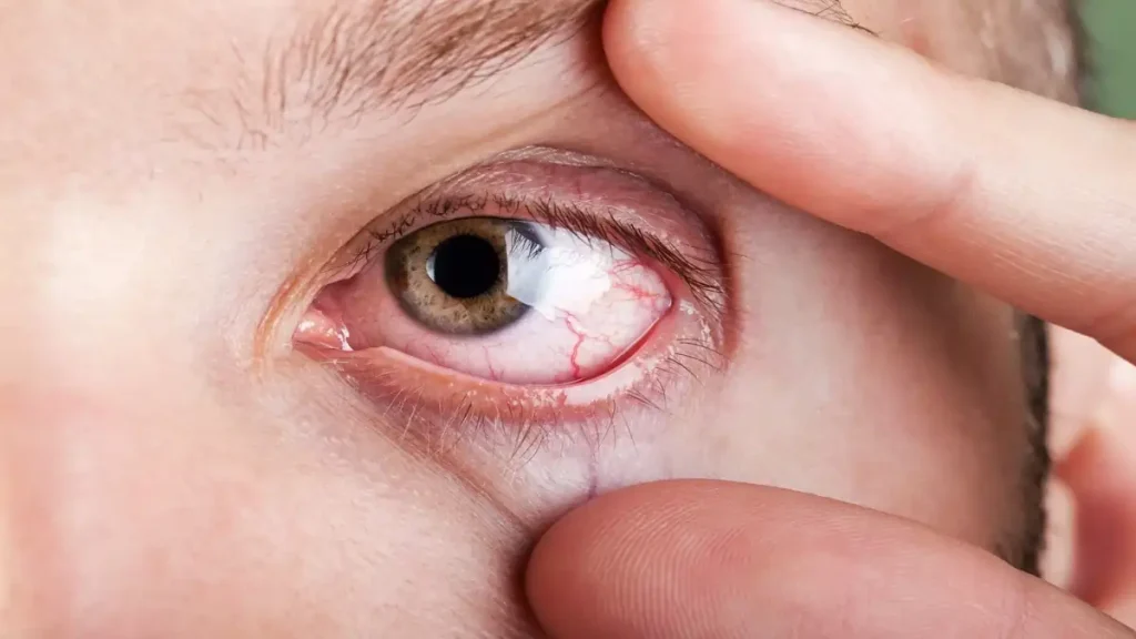
Due to the proximity of the orbital floor to the eye and its structures, ophthalmologic evaluation is crucial in the initial assessment of orbital floor fractures. A thorough examination of visual acuity, eye movements, pupillary reactions, and intraocular pressure should be performed to assess for any damage to the eye. Additionally, a detailed ophthalmologic examination can identify potential complications, such as retinal detachment or optic nerve injury, which may require immediate intervention.

Operative management is often necessary for orbital floor fractures, especially in cases of significant displacement or entrapment of the orbital contents. The decision for surgery is based on the severity of symptoms, radiographic findings, and ophthalmologic evaluation. Repairing the orbital floor not only helps restore normal eye function but also prevents long-term complications, such as chronic diplopia or enophthalmos.

Medial Orbital Wall Fracture

When approaching a medial orbital wall fracture, it is important to consider the surgical approach that will effectively restore orbital function and minimize complications. The transcaruncular incision is a commonly used approach due to its aesthetic advantage as it hides scars within the natural creases of the eye. However, there are several risks associated with this approach that must be carefully addressed.

One potential risk is injury to the inferior oblique muscle and adjacent neurovascular structures. The surgeon must be cautious during dissection to avoid damaging these important structures.

Medial Orbital Wall Fracture

Additionally, meticulous hemostasis should be ensured to prevent excessive bleeding that may compromise visualization and increase the risk of injury.

Plate placement over the posterior ledge is of utmost importance in repairing a medial orbital wall fracture. The posterior ledge provides stability and support to the orbital volume, and placing a plate over it helps restore and maintain the proper volume. For larger defects, the use of preformed, contoured orbital plates can be beneficial. These plates are specifically designed to fit the anatomical contours of the orbit, resulting in a secure and precise reconstruction.

Naso-Orbito-Ethmoid Fractures

Naso-Orbito-Ethmoid (NOE) fractures are a type of facial fracture that involve the bones of the nose, orbit, and ethmoid complex. These fractures are complex in nature due to the involvement of multiple structures.

Signs and symptoms of NOE fractures can vary depending on the severity of the injury. Common manifestations include swelling and deformity of the nose, bruising around the eyes, double vision, and difficulty breathing through the nose. In severe cases, patients may experience cerebrospinal fluid leakage from the nose or loss of vision.

Treatment approaches for NOE fractures typically involve surgical intervention. The main goal is to restore normal facial appearance and function. One common surgical technique used for NOE fractures is the Modified Cantor’s Transcaruncular (MCT) approach. This approach provides access to the central fragment, which is the key component of the fracture. It allows for precise reduction and fixation of the fractured bones, facilitating proper alignment and healing.

Overall, Naso-Orbito-Ethmoid fractures are complex injuries that require specialized surgical management. Prompt identification and treatment are crucial to prevent long-term complications and achieve optimal outcomes. By understanding the signs and symptoms of NOE fractures and utilizing appropriate surgical approaches, healthcare professionals can provide efficient and effective care to individuals with these injuries.

How is Orbital Reconstruction Performed?

Orbital reconstruction is a surgical procedure that involves restoring the normal appearance and function of the eye socket, or orbit, which may have been damaged due to trauma or disease. A complex and delicate operation, orbital reconstruction requires a multidisciplinary approach involving various specialists, such as oculofacial plastic surgeon and maxillofacial surgeons. This procedure aims to not only repair the structural integrity of the orbit but also to address any functional deficits, including improvement of vision and eye movement.

The process of orbital reconstruction involves several key steps, including the preoperative assessment, surgical planning, and the actual reconstructive surgery. Through meticulous planning and careful execution, orbital reconstruction can successfully restore the patient’s quality of life and aesthetic appearance.

Conservative Management

Conservative Management for Orbital Reconstruction (1)

Conservative Management Approach for Chronic Back Pain:

The conservative management approach for chronic back pain focuses on the use of non-invasive treatments, lifestyle modifications, and monitoring methods to alleviate pain and improve the patient’s quality of life.

Non-invasive treatments play a crucial role in managing chronic back pain. These may include physical therapy, which involves exercises and stretches to strengthen the muscles supporting the spine and improve flexibility.

Additionally, non-steroidal anti-inflammatory drugs (NSAIDs) are commonly prescribed to reduce inflammation and pain. Moreover, the use of heat or cold therapy, such as applying hot or cold packs to the affected area, can provide temporary relief. In some cases, transcutaneous electrical nerve stimulation (TENS) may be used to disrupt pain signals.

Lifestyle modifications are an integral part of conservative management for chronic back pain. Patients are encouraged to maintain a healthy weight to reduce strain on the spine. Regular exercise, such as walking, swimming, or yoga, can help strengthen core muscles and improve posture. Ergonomic adjustments, such as using proper lumbar support chairs and avoiding prolonged sitting or heavy lifting, are also recommended. Furthermore, stress management techniques, including relaxation exercises and cognitive-behavioral therapy, can help alleviate the psychological impact of chronic back pain.

Monitoring methods are essential to track the progress and effectiveness of conservative management for chronic back pain. Regular follow-up visits with healthcare professionals allow for adjustments to treatment plans as needed. Patient-reported outcomes, such as pain scales and functional assessments, provide valuable insight into the effectiveness of conservative management strategies. In some cases, imaging studies, such as X-rays or MRI scans, may be used to identify any structural abnormalities or assess the progression of the condition.

Use of Implants

Orbital implants support the natural volume of the eye socket after enucleation or evisceration surgeries. Various materials can be used for orbital implants, each with its own set of advantages and disadvantages.

Bone is a traditional material used for orbital implants. It offers excellent support and osteointegration, as it can allow the patient’s own bone to grow into the implant. However, bone implants can be limited by their availability and malleability. Harvesting bone from the patient’s own body can cause additional surgical sites and potential complications.

Use of Implants For Eyes

Cartilage implants, derived from the patient’s rib or nasal septum, provide a natural feel and appearance. They are malleable and can be easily shaped during the surgery for a custom fit. However, cartilage has limited availability and a risk of resorption over time.

Medpor, a porous polyethylene material, is readily available and offers good support. It is malleable and can be easily carved to achieve desired contours. However, Medpor implants are not osteointegrative, and there is a risk of migration and exposure through the skin.

Titanium implants are durable, inert, and readily available. They provide good support and do not require removal unless there are complications. However, titanium implants are not malleable and require preoperative customization. Also, they can be expensive.

Poly(L-lactide) and polydioxanone implants are biodegradable and have the advantage of being absorbed by the body over time. They offer good malleability and can be easily shaped during surgery. However, they have limited availability and a risk of complications such as infection and migration.

Open Reduction Internal Fixation

Open Reduction Internal Fixation (ORIF) is a surgical technique used to treat fractures by aligning and fixing the fractured bones using screws, plates, or rods. The procedure involves making an incision near the fractured site to gain direct access to the bone.

First, the surgeon gently manipulates the fragments into their normal anatomical alignment. This ensures proper healing and allows for the best possible recovery. Once the alignment is achieved, the surgeon then uses screws, plates, or rods to hold the fractured bones in place. These devices provide stability and support to the bone, allowing for early mobilization and better healing.

ORIF offers several advantages over other methods of fracture treatment, such as traction or closed reduction. Traction is a non-surgical method that involves using a pulling force to align the bones. However, it often fails to achieve accurate alignment and may result in prolonged immobilization. On the other hand, closed reduction may be successful in aligning the bones but lacks the stability provided by surgical fixation.

The use of screws, plates, or rods in ORIF ensures that the fractured bones remain in their correct position during the healing process. This stability allows for early mobilization and can help reduce the risk of complications, such as non-union or malunion. Moreover, ORIF allows for more precise alignment of the bones, which is crucial for optimal healing and functional recovery.

Orbital Reconstruction Outcomes

Orbital reconstruction is a surgical procedure used to restore the form and function of the orbit, which is the bony cavity that houses the eye. The outcomes of orbital reconstruction are aimed at achieving optimal aesthetic appearance and preserving vision.

Successful orbital reconstruction outcomes depend on several factors. First, atraumatic technique is crucial to minimize tissue damage during the surgery. This includes careful handling of the tissues and meticulous dissection to preserve blood supply. Anatomical reduction is another key factor, as it involves restoring the bones and soft tissues to their pre-injury position. This is essential for achieving proper orbital volume and contour.

Stable fixation is also vital for successful orbital reconstruction outcomes. It involves securing the bones in their correct position using plates, screws, or other implants. Stable fixation ensures long-term support and promotes proper healing.

Complications can arise from orbital trauma and they can have a significant impact on both form and function. These complications include diplopia (double vision), enophthalmos (sinking of the eye into the socket), ectropion (outward turning of the eyelid), and infraorbital nerve injury, among others. These complications can lead to both aesthetic and functional impairment, affecting the patient’s ability to see properly and causing discomfort.

Computer-assisted planning and surgery play a crucial role in improving postoperative results in orbital reconstruction. These technologies help in preoperative planning, allowing surgeons to accurately assess and manipulate the virtual model of the orbit before the actual surgery. This enhances the precision and predictability of the surgical procedure, resulting in improved outcomes and patient satisfaction.

Orbital Reconstruction Summary

In conclusion, orbital reconstruction is a complex surgical procedure used to restore the normal anatomy and function of the orbit (eye socket) following trauma or disease. Computer-assisted planning and intraoperative navigation/imaging have become valuable tools in planning and performing these surgeries.

Computer-assisted planning involves the use of advanced imaging techniques, such as CT scans or MRI, to create a three-dimensional model of the patient’s orbit. This model enables the surgeon to accurately analyze the extent of the injury or deformity, plan the surgical approach, and simulate potential outcomes.

Intraoperative navigation and imaging technologies, such as stereotactic systems or real-time fluoroscopy, assist the surgeon in accurately placing surgical instruments and implants during the procedure. These technologies provide real-time feedback on the position and alignment of the reconstructive components, ensuring optimal surgical results.

Secondary orbital reconstructions are indicated in cases where the initial reconstruction has failed or when new deformities or functional deficits develop over time. Potential complications of orbital trauma include enophthalmos (sunken eyeball), diplopia (double vision), ocular motility disturbances, and aesthetic deformities. These complications may require additional surgeries to correct or improve the initial reconstruction.

Internal orbital reconstruction refers to the restoration of the pretrauma anatomy using autologous bone or synthetic implants. The challenges associated with this procedure include achieving precise alignment and stability of the reconstructed orbit, preserving lacrimal system function, and avoiding injury to important structures, such as the optic nerve and vessels.

If you have any questions regarding orbital reconstruction, we encourage you to contact Dr. Bidar for your confidential consultation.

CONTACT US